Search history
Clear allSearch by image
XDrag and drop an image here or upload an image
Max 5MB per image
UploadSign In | Join
Search history
Clear allSearch by image
XDrag and drop an image here or upload an image
Max 5MB per image
UploadSign In | Join
X Email Mobile
Yiwu Conor medical equipment Co., LTD 2yr.
Contacts Amina Chat
Mobile 86-15058651598
E-mail 785069582@qq.com


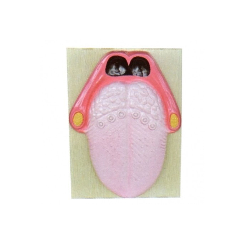
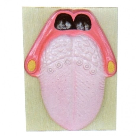
BM1088 taste bud model is developed to meet the needs of medical colleges, health schools, and hospital clinical treatment teaching. It has strong scientific properties, is easy to operate, is convenient for students to understand, has practical value for teaching, and is reasonably priced. Specification: 36x27x6cm
Taste bud model shows structural morphology.
Taste buds are taste receptors, mainly distributed on the back of the tongue, especially at the tip and sides. They are oval-shaped small bodies located in the epithelium, about 80μm long and 40μm thick. They are mainly distributed in the side wall epithelium of the papillae near the groove, and they can also be seen in the epithelium of other places such as fungus-like papillae, soft palate, and epiglottis. Taste buds are special structures differentiated from the epithelium. Their base is located above the basal membrane, and they are covered by keratinocytes on the surface, with a central hole, which is the taste hole, leading to the oral cavity. Under the light microscope, there are two types of cells that constitute taste buds, namely bright cells and dark cells, with the former being larger and the latter being slender. The long axis of the cells is perpendicular to the epithelial surface. Near the taste hole, the top of the cells has finger-like cytoplasmic projections called taste hairs.
Update time:


TOP