
Search history
Clear allSearch by image
XDrag and drop an image here or upload an image
Max 5MB per image
UploadSign In | Join

Search history
Clear allSearch by image
XDrag and drop an image here or upload an image
Max 5MB per image
UploadSign In | Join
Yiwu Conor medical equipment Co., LTD 2yr.
Contacts Amina Chat
Mobile 15058651598
C19519879654

WhatsApp 19519879654
Our company isCollection Self-service R&D, production, sales, and serviceA comprehensive high-tech enterprise integrated as one, with a complete and scientific quality management system.
The main products include high-quality onesHuman skeletal structure, anatomy, oral teaching models, and cardiopulmonary resuscitation simulators; first aid skill training, nursing skill training, comprehensive specialty training, women and infant care skill training, traditional Chinese medicine acupunctureMedical model and other skill training experimental system models.
Testicle enlarged model, anatomical model of the male reproductive system, medical teaching model
Testicle model
Structural features:The model is designed using a 3.5-fold enlarged normal male testicle. It displays the inner side and cross-section of the testicle, providing detailed information on its internal structures, such as efferent ducts, tunica albuginea, tunica vaginalis cavity, visceral layer of the tunica vaginalis, seminiferous ducts, vas deferens, and the testicular network. This is a valuable product for reproductive science and urology education. It is widely used in various urology hospitals as a primary tool for patient-doctor communication, and is also suitable for educational demonstrations in hospitals. The upper half of the model shows a cross-section of the testicle, highlighting its external and internal structures.
Size:10 times enlarged, 11x11x26 cm, Weight: 270 G
Material:Imported PVC materials, imported paints, computer color matching, painting
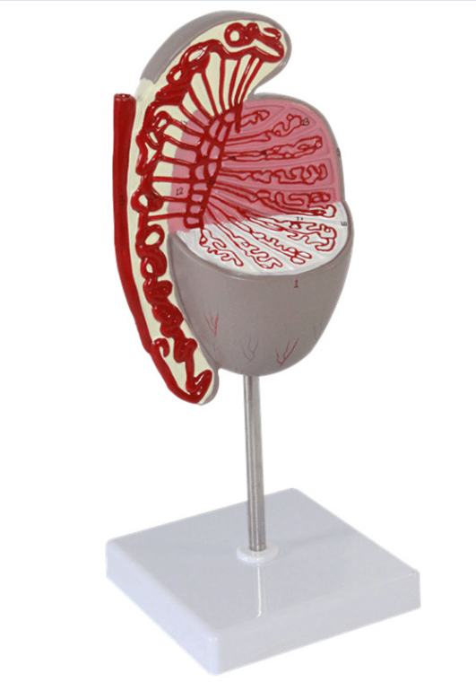
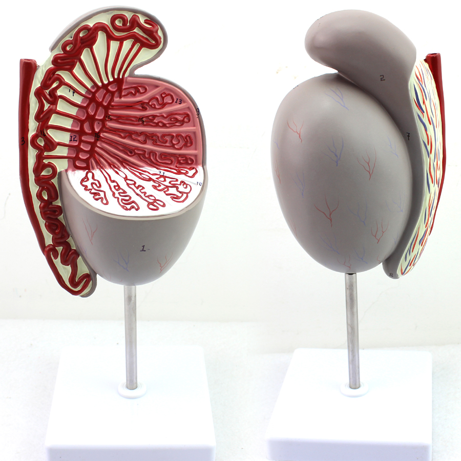
The testes are located within the scrotum, one on each side. Generally, the left testicle is slightly lower than the right by 1 cm. The testes are slightly flattened and oval in shape, with a smooth surface. They consist of an inner and an outer surface, as well as anterior, posterior edges, and upper and lower ends. The anterior edge is free; the posterior edge has blood vessels, nerves, and lymphatic vessels entering and exiting, and it contacts the epididymis and the testicular part of the vas deferens. The upper end and posterior edge are attached to the head of the epididymis, while the lower end is free. The outer surface is more convex, whereas the inner surface is more flat.
Structure of the testicles
The fibrous membrane, known as the white membrane, thickens along the posterior edge of the testicle and protrudes into the testicle to form the testicular mediastinum. Many connective tissue septa emanate from the mediastinum, dividing the testicular parenchyma into numerous testicular lobules. Within the testicular lobules, coiled seminiferous tubules are present, and the epithelium of these tubules produces sperm. The connective tissue between the tubules contains interstitial cells that secrete male hormones. The seminiferous tubules combine to form direct seminiferous ducts, which enter the testicular mediastinum and interweave to form the testicular network. One to five testicular efferent ducts emerge from the testicular network and pass above the posterior edge of the testicle into the epididymis.
Update time:


TOP


