Search history
Clear allSearch by image
XDrag and drop an image here or upload an image
Max 5MB per image
UploadSign In | Join

Search history
Clear allSearch by image
XDrag and drop an image here or upload an image
Max 5MB per image
UploadSign In | Join
Yiwu Conor medical equipment Co., LTD 2yr.
Contacts Amina Chat
Mobile 15058651598
C19519879654

WhatsApp 19519879654
Function Description:
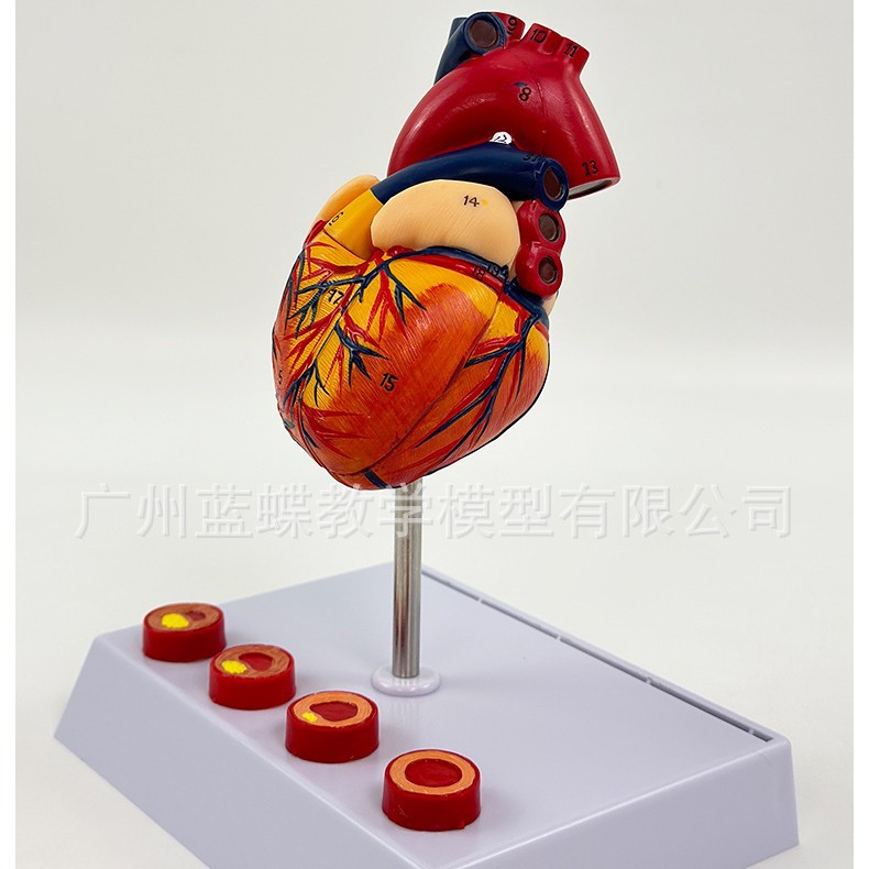
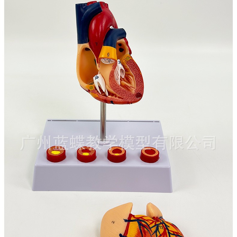
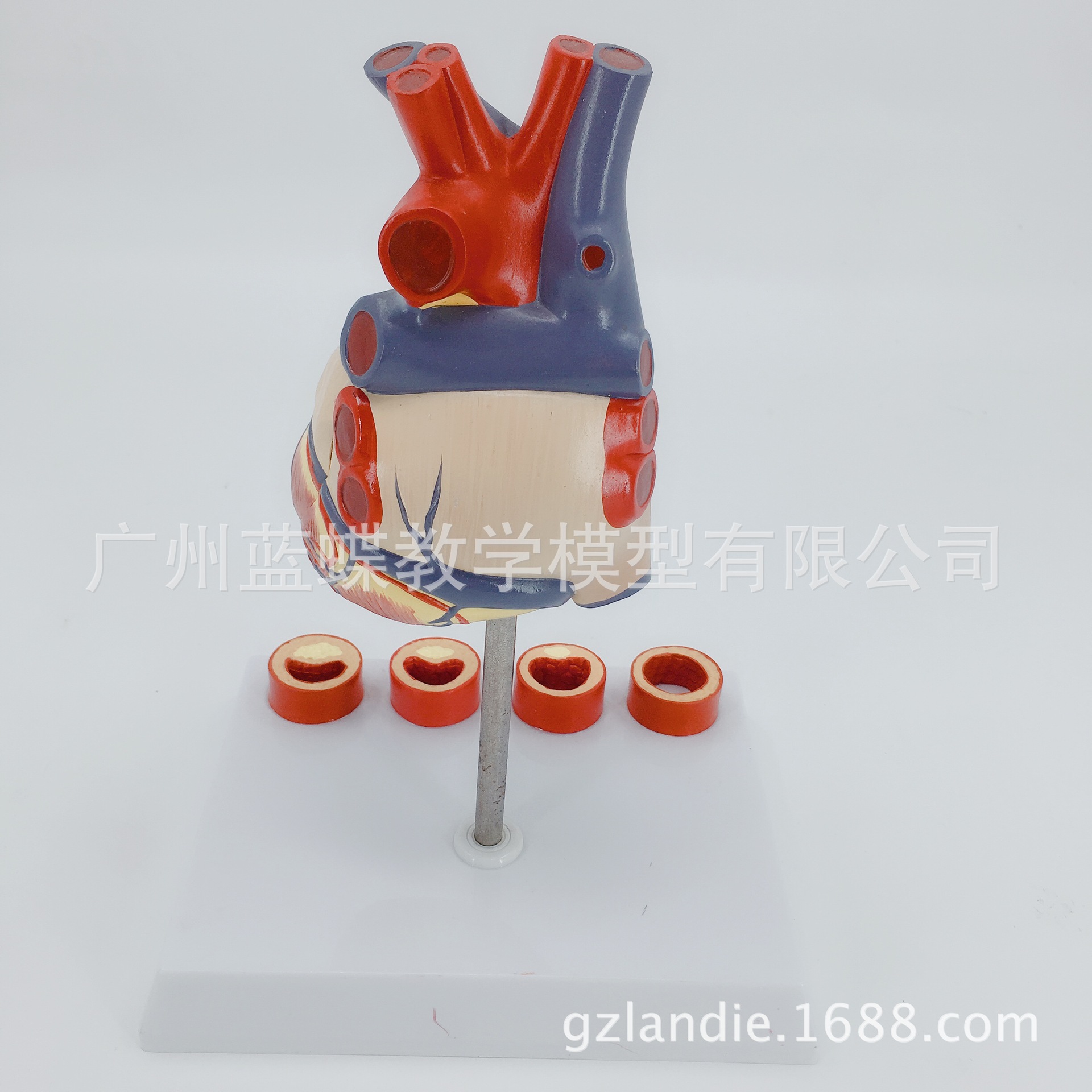
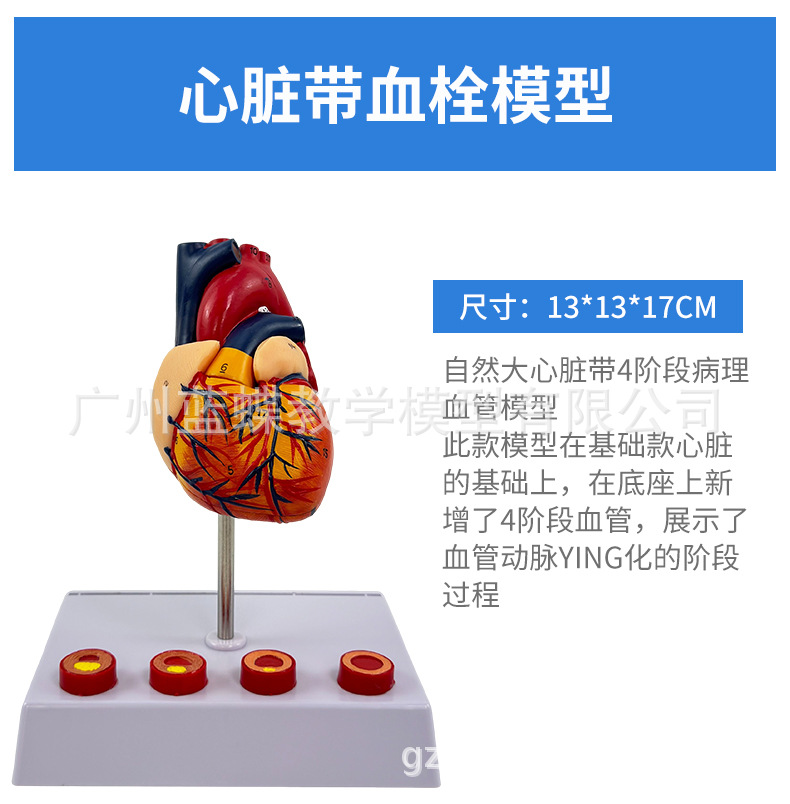
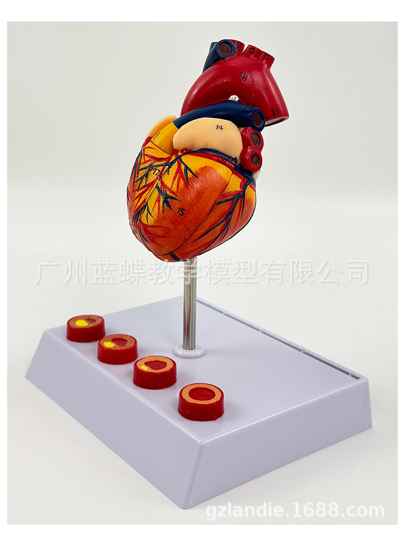
The human heart consists of two components that illustrate the normal structural arrangement of the heart, including arteries, veins, and valves. It is equipped with a four-stage blood clot formation demonstration model, making it particularly suitable for medical education and patient-doctor communication.
Model Structure: The model depicts an adult heart at approximately 1:1 scale, in a relaxed state. Placed on a stent according to the normal physiological position of the human body. A transverse cut along the root of the pulmonary artery shows the internal structure of the left and right atria and the pulmonary artery, including the pulmonary semilunar valve. A longitudinal oblique cut along the atria reveals the internal structure of the left and right ventricles. The right atrium displays the superior and inferior vena cava openings, inferior vena cava valve, coronary sinus opening and valve, papillary notch, right atrioventricular orifice; the right atrium also shows the left and right pulmonary venous orifices and the left atrioventricular orifice. The right ventricle features the superior ventricular crest, tricuspid valve, arterial cone, cord, papillary muscles, trabeculae, and pulmonary valve; the left ventricle shows the mitral valve, chorda tendineae, papillary muscles, trabeculae, and aortic valve. The atrial septum and ventricular septum are displayed. The external morphology of the heart, associated blood vessels, and branches are shown. The tube-like structure, interventricular sulci, and auriculars, myocardial fibers, cardiac vortex, cardiac notches, fat, and arterial ligaments are presented. The left and right coronary arteries and their branches are shown; the coronary sinus and its branches are depicted; branches originating from the ascending aorta and aortic arch are illustrated; the superior vena cava, inferior vena cava, and their branches are presented; the pulmonary veins and pulmonary artery are displayed. The walls of the left and right ventricles of the model, as well as the surface cut along the arterial pulse, are detachable.
Thrombotic process model: The process in which blood coagulates or certain components in the blood aggregate together within the heart or vascular lumen of a living organism to form solid masses is called thrombosis. The solid masses formed during this process are referred to as thrombi.

Update time:


TOP