Search history
Clear allSearch by image
XDrag and drop an image here or upload an image
Max 5MB per image
UploadSign In | Join

Search history
Clear allSearch by image
XDrag and drop an image here or upload an image
Max 5MB per image
UploadSign In | Join
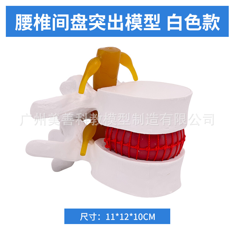
 lumbar vertebra blood cell-white
腰椎血球-白色款 |
CN¥ 0.0 |
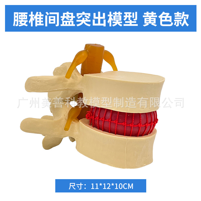
 lumbar vertebra blood cell – yellow model
腰椎血球-黄色款 |
CN¥ 0.0 |
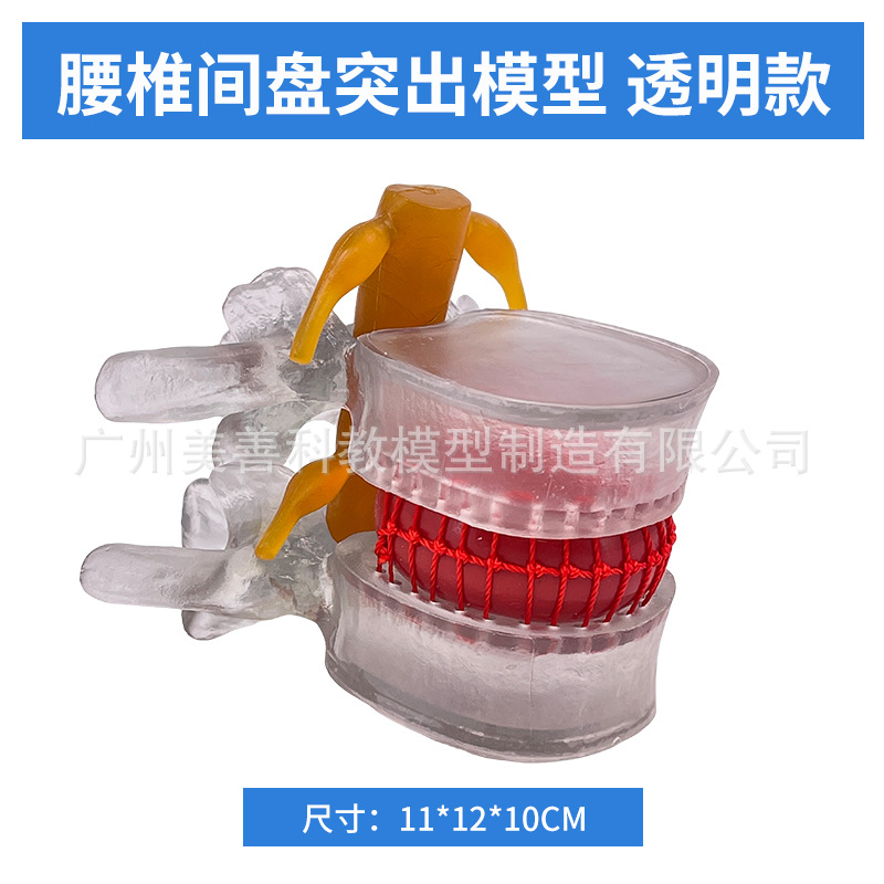
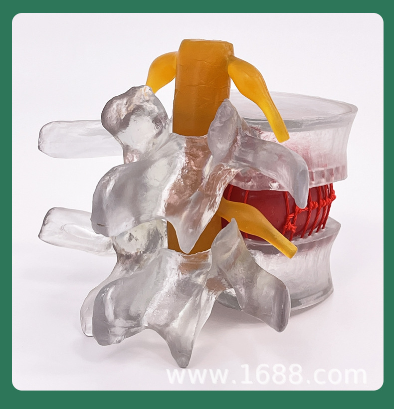
 lumbar vertebral blood cell – transparent style
腰椎血球-透明款 |
CN¥ 0.0 |
Global Commodity Supplier 1yr.
Contacts Wei Fengwei Chat
Mobile 15356982475
WeChat 15356982475
.....................................................................................................................................................
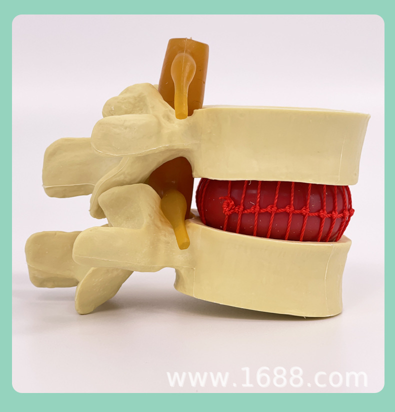
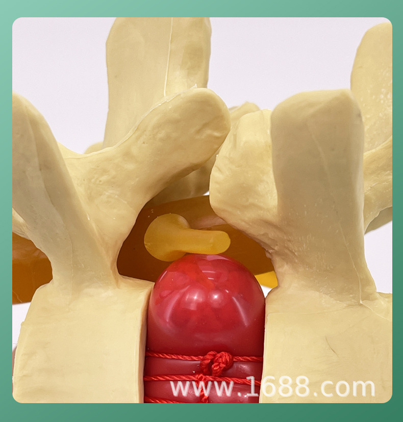
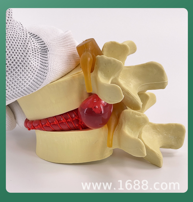
Lumbar disc herniation / Demonstration model
Lumbar disc herniation is one of the more common conditions. This occurs due to degenerative changes in various parts of the lumbar disc—such as the nucleus pulposus, annulus fibrosus, and cartilaginous layers—especially the nucleus pulposus. Under the influence of external forces, the annulus fibrosus ruptures, and the nucleus pulposus protrudes from the rupture site into the back or spinal canal, causing irritation or compression of adjacent spinal nerve roots. This leads to lower back pain, numbness, and pain in one or both lower limbs, among other clinical symptoms. Lumbar disc herniation is most prevalent at the L4-L5 and L5-S1 levels, accounting for approximately 95% of cases.
In this context, we developed this model
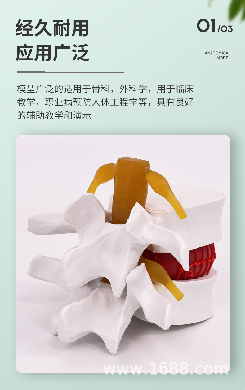
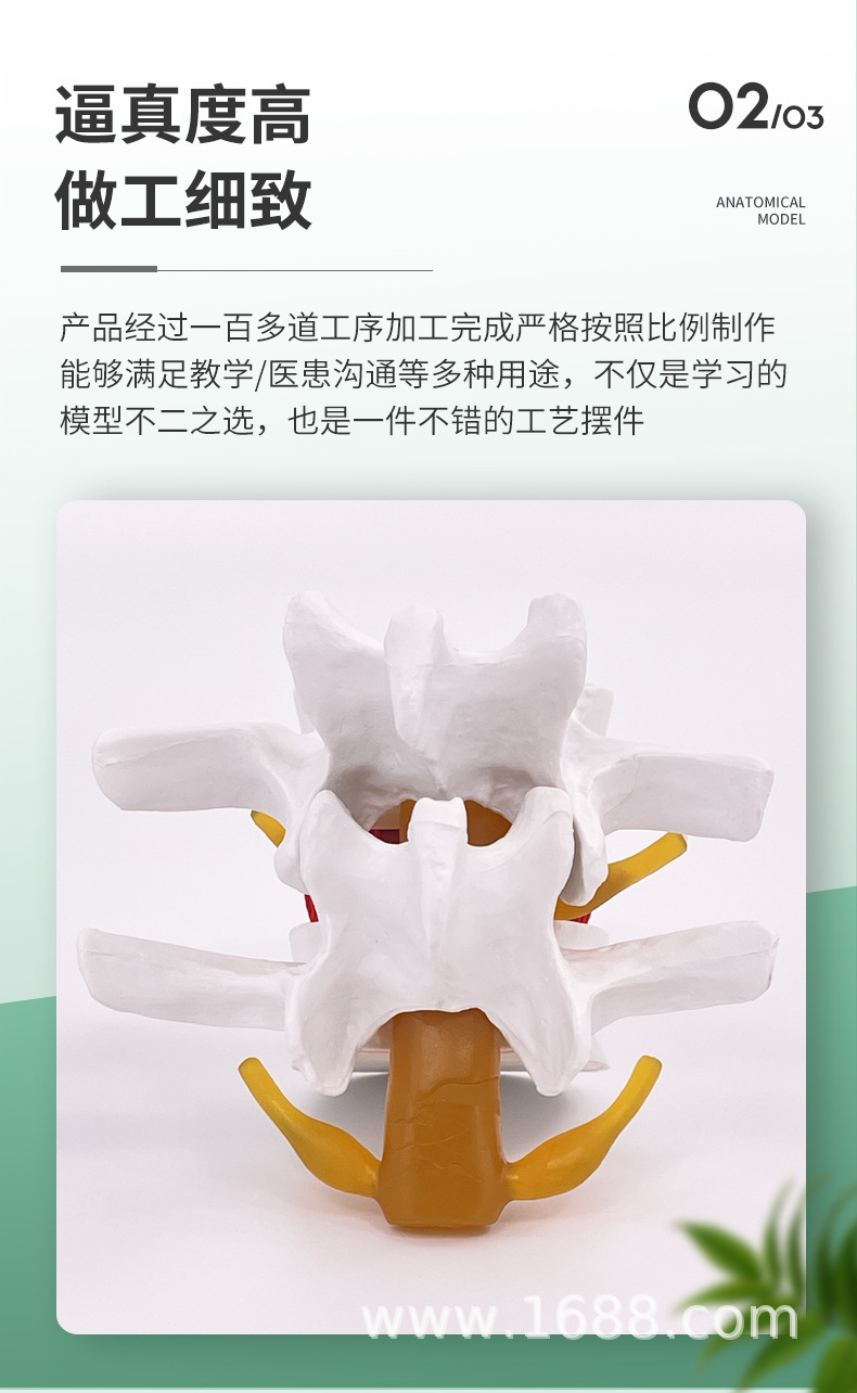
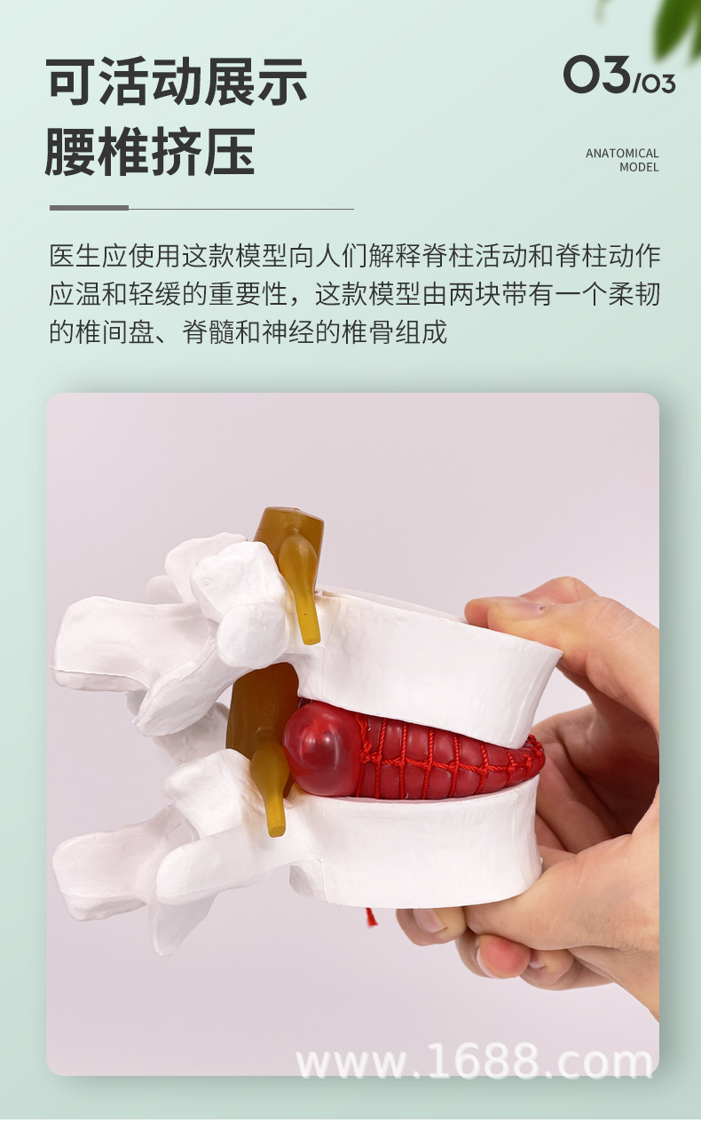
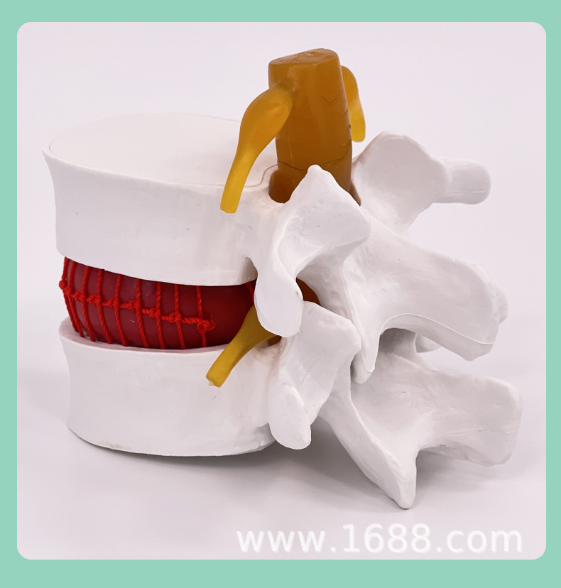
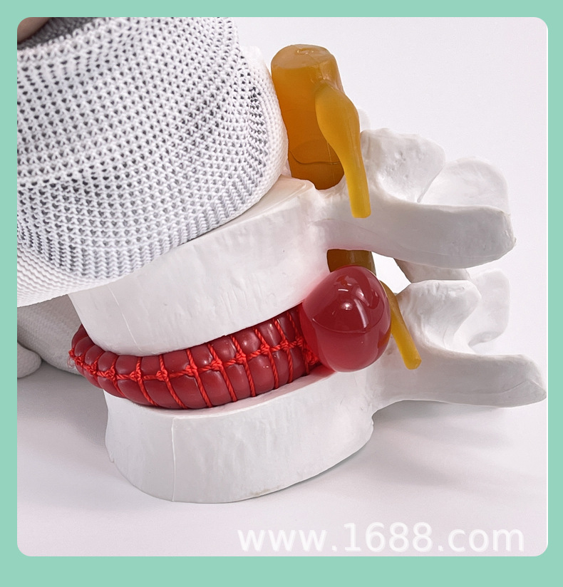
This model features the fourth and fifth lumbar vertebrae enlarged by 1.5 times, consisting of intervertebral discs and spinal nerves. The intervertebral discs are made of a soft elastic material to facilitate user demonstrations. It also shows the changes that occur in the intervertebral discs when the body bends or curls.
This model is widely applicable in orthopedics, used for clinical teaching, occupational disease prevention, ergonomics, sports education, and more. It serves as a valuable aid for teaching and demonstration!










Update time:


TOP